2. Perio Anatomy
Aug 19, 2020 ·
6m 35s
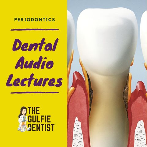
PERIODONTIUM 1 Gingiva 2 Attachment apparatus* a. Pdl b. Alveolar bone c. Cementum (has dead cells) acellular cementum PERIODONTAL TISSUE : (has living cells) a) Gingiva b) Periodontal ligament c)...
show more
PERIODONTIUM
1 Gingiva
2 Attachment apparatus*
a. Pdl
b. Alveolar bone
c. Cementum (has dead cells) acellular cementum
PERIODONTAL TISSUE : (has living cells)
a) Gingiva
b) Periodontal ligament
c) Alveolar bone
PARTS OF GINGIVA
Normal range of gingival sulcus depth is- 2-3mm
Colour of normal gingiva is an interplay between – keratin layer, melanine, blood vessels, epithelial thickness.**
FREE GINGIVA – Also known as unattached / marginal gingiva. From the gingival margin till the free gingival groove / base of the sulcus.
(sulcus is in healthy gums, whereas pocket is in unhealthy / diseased
gums) KERATINIZED
ATTACHED GINGIVA- From free gingival groove (base of the sulcus) to the
mucogingival junction. KERATIINIZED
o Healthy one shows stippling.
o Best views by drying the gingiva.
o Highest width is seen in incisors-
Maxillary : 3.4-4.5
Mand - 3.3-3.9**
o Narrowest seen in molars
Max 1.9mm
Mand 1.8mm
ALVEOLAR MUCOSA
o From mucogingival junc to fold
o Non keratinized
INTERDENTAL GINGIVA
a) Anterior – pyramidal
b) Posterior — col shape
c) Midline diastema — triangular
FREE GINGIVAL GROOVE
MUCOGINGIVAL JUNCTION
BIOLOGICAL WIDTH
Biological width — junctional epithelium + connective tissue = 2 mm***
show less
1 Gingiva
2 Attachment apparatus*
a. Pdl
b. Alveolar bone
c. Cementum (has dead cells) acellular cementum
PERIODONTAL TISSUE : (has living cells)
a) Gingiva
b) Periodontal ligament
c) Alveolar bone
PARTS OF GINGIVA
Normal range of gingival sulcus depth is- 2-3mm
Colour of normal gingiva is an interplay between – keratin layer, melanine, blood vessels, epithelial thickness.**
FREE GINGIVA – Also known as unattached / marginal gingiva. From the gingival margin till the free gingival groove / base of the sulcus.
(sulcus is in healthy gums, whereas pocket is in unhealthy / diseased
gums) KERATINIZED
ATTACHED GINGIVA- From free gingival groove (base of the sulcus) to the
mucogingival junction. KERATIINIZED
o Healthy one shows stippling.
o Best views by drying the gingiva.
o Highest width is seen in incisors-
Maxillary : 3.4-4.5
Mand - 3.3-3.9**
o Narrowest seen in molars
Max 1.9mm
Mand 1.8mm
ALVEOLAR MUCOSA
o From mucogingival junc to fold
o Non keratinized
INTERDENTAL GINGIVA
a) Anterior – pyramidal
b) Posterior — col shape
c) Midline diastema — triangular
FREE GINGIVAL GROOVE
MUCOGINGIVAL JUNCTION
BIOLOGICAL WIDTH
Biological width — junctional epithelium + connective tissue = 2 mm***
Information
| Author | Dr.Mayakha Mariam |
| Website | - |
| Tags |
-
|
Copyright 2024 - Spreaker Inc. an iHeartMedia Company
